- Home
- > Nature online
- > The science of natural history
- > The scientific process
- > Methods for examining specimens
Primary navigation
Methods for examining specimens
Scientists use different methods to examine specimens. Lots of detail can be seen with the naked eye but new technologies have opened up lots of other possibilities as well.
Microscopes have become more powerful, allowing scientists to examine specimens in ever-greater detail, and X-rays allow them to look inside a specimen at the bone structure.
At the other end of the scale, satellite images enable scientists to see distances of hundreds of kilometres in a single picture, which is useful when studying large-scale processes like hurricanes.

Satellite images
Images that are 10s or 100s of kilometres across can show how a habitat changes in relation to geography and soil composition. They are also used to track changes over time, for example, tracking the recovery of forests after being cleared.
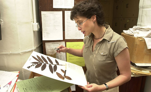
The naked eye
The naked eye can see a vast amount of detail in a specimen. Coupled with a hand lens, it is the day-to-day way of examining specimens used by our scientists. A great deal of taxonomic work requires no complex technology, just a trained eye.
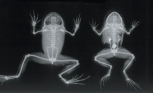
X-rays
X-rays don't magnify a specimen but they allow scientists to see the internal detail without dissecting it. These X-rays are made from frogs looked after in the Museum's collections. The picture reveals the internal bone structures, which help to distinguish many species of fish and amphibian.
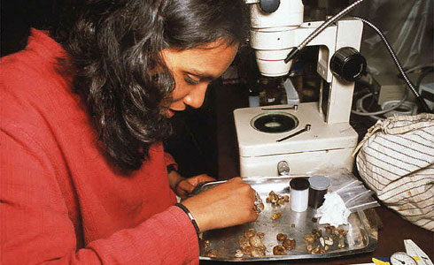
Light microscopes
Most laboratories contain a basic light microscope, used for examining a specimen in detail and aiding dissections and preparations.
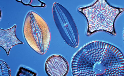
The power of light microscopes
Above is a group of fossil diatoms from Hungary viewed through a light microscope. Most diatoms are single-celled organisms but they can be seen in detail at this level of magnification.
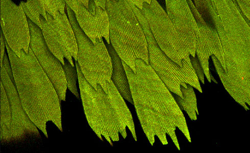
Confocal microscopes
A blue morpho butterfly seen through a confocal microscope. Confocal microscopes can be used to increase contrasts of colour or to create 3-dimensional images.
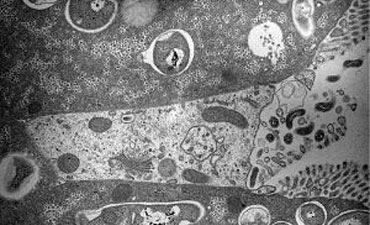
Transmission electron microscopes
Transmission electron microscopes can find fine detail inside a specimen. By magnifying organisms more than 100,000 times, they allow us to look at the shape and structure of cells, as shown above.
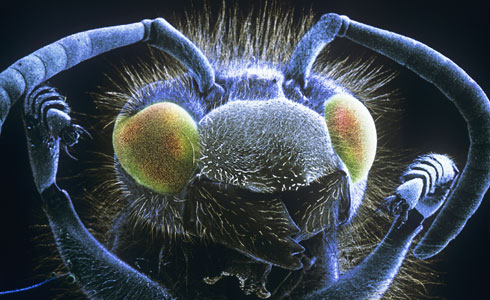
Scanning electron microscopes
This is a common wasp seen through a scanning electron microscope. Scanning electron microscopes can magnify objects up to 200,000 times. It makes tiny details visible, such as pollen grains or the structures of insect mouth parts. This makes it easier to categorise species.
Related information
Toolbox
Art, nature and imaging

Discover how natural history art and imaging techniques have developed since the 17th century and explore selected artworks from the Museum’s world-class art collections.
- Contact and enquiries
- Accessibility
- Site map
- Website terms of use
- © The Trustees of the Natural History Museum, London
- Information about cookies
- Mobile