Under the microscope
To understand what a meteorite is made of, and what has happened to it throughout its life, it has to be studied in minute detail. This is done with the help of microscopes.
Optical microscopes
Optical microscopes are the simplest type of microscope. They have been used to study rocks, including meteorites, for a long time. The meteorite needs to be cut so thinly that light can pass through. For most minerals this is about 0.03 millimetres thick, as thin as a pencil line. The optical microscopes magnify the light that passes through the meteorite, allowing tiny crystals of mineral to be seen and studied.
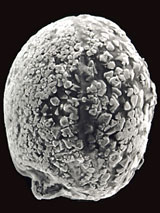
An electron image of a micrometeorite.
Electron microscopes
Microscopes known as scanning electron microscopes (SEM) allow minerals 0.001 millimetres thick to be studied. The SEMs work by firing a beam of electrons at the meteorite. This causes the elements within the minerals to emit X-rays. Each element produces a slightly different X-ray, allowing it to be identified. The X-rays can also be used to trace how the elements that make up minerals have changed through chemical reactions. By knowing what chemical reactions have taken place, meteoriticists can work out what has happened throughout a meteorite's life.
The most powerful microscope used to study meteorites is called the transmission election microscope (TEM). TEMs can pass a beam of electrons, tiny sub-atomic particles, through a section of meteorite only 600 atoms thick. They are so powerful that they can be used to examine the atomic structure of minerals, and can even see irregularities caused by radiation and impacts.
Toolbox

In 2003 nearly 12,000 scientists from over 60 countries came to work at the Museum.