Pseudomicrothorax dubius (ciliate)
Watch the protozoan Pseudomicrothorax dubius ingesting the blue-green algae, or cyanobacterium, Nostoc muscorum. © Agnieszka Pajdak-Stós, Institute of Environmental Sciences, Jagiellonian University, Poland, www.eko.uj.edu.pl
Pseudomicrothorax dubius is a common freshwater and terrestrial ciliated protozoan (ciliate).
It specialises in feeding on filamentous prey, especially cyanobacteria. The ingested cyanobacteria give P. dubius its green to blue-green colour.
Find out more about cyanobacteriaA biological control agent?
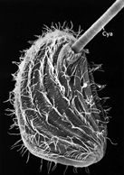
A scanning electron micrograph showing P. dubius ingesting a cyanobacteria (Cya) © Elsevier. From Hausmann & Peck (1979)
Cyanobacteria occur worldwide, especially in calm, nutrient-rich waters. Some cyanobacterial species produce toxins that affect animals and humans, and people can be exposed by drinking or bathing in contaminated water. The most frequent and serious health effects are caused by drinking water containing the toxins. Swimmers may also suffer allergic reactions such as eye irritation and blisters caused by contact with the toxins.
A possible way to eliminate harmful cyanobacteria is to use biological control agents. Ciliate grazers such as P. dubius have been suggested as possible candidates and their potential has been demonstrated in the laboratory. Currently, however, the logistical problems of producing and storing sufficient amounts of ciliate grazers prevent their effective use for the control of cyanobacteria in large water bodies such as lakes and reservoirs (Sigee et al., 1999).
-

Taxonomy
Find out about the taxonomy of this fascinating ciliate.
-

Distribution
Discover the habitats, distribution and dispersal mechanisms of P. dubius and how, thanks to its ability to thrive in particular environmental conditions, it has potential as a biological indicator of pollution.
-

Biology
Learn about the biology of P. dubius, including the organism's morphology, feeding habits, cell structure and method of reproduction.
-

Behaviour
Find out about the behaviour of P. dubius and how certain cyanobacteria defend themselves against its attack.
-

References
Get information about referenced journal publications.
Images

P. dubius viewed under light microscopy.Green colour is due to ingested cyanobacteria. © William Bourland

P. dubius showing the trichocysts on the cell periphery and cytopharyngeal basket © DJ Patterson

Scanning electron micrograph of P. dubius. The cell is on its side with its anterior end on the right. The arrow shows the position of the mouth. © University of Heidelberg and Land Oberosterrich des OO. Landesmuseums.

Pseudomicrothorax dubius, silverstained cell showing the three adoral membranelles, the rows of somatic cilia, the sausage-shaped macronucleus and the spherical micronucleus. © William Bourland
P. dubius cells and a cyst (indicated by the arrow) © Agnieszka Pajdak-Stós, Institute of Environmental Sciences, Jagiellonian University, Poland, www.eko.uj.edu.pl

Pseudomicrothorax dubius with numerous endosymbiotic cells of 'Candidataus Cryptoprodotis polytropus', detected through fluorescence in situ hybridisation (FISH). Ferrantini et al. (2009) Journal of Eukaryotic Microbiology 56(2): 119-129. Published online: 3 March 2009. DOI: 10.1111/j.1550-7408.2008.00377 © Wiley-Blackwell.

Ventral view of Pseudomicrothorax agilis. © William Bourland

A scanning electron micrograph showing P. dubius ingesting a cyanobacteria (Cya). © Elsevier
About the author

Dr Alan Warren
Researching the systematics of ciliated protozoa (ciliates) biodiversity of marine ciliates and the use of ciliates as bioindicators of environmental quality.
Toolbox
Share this
Glossary
Ciliate
A protozoan that has cilia (specialised cell structures that look like hair- or tail-like projections).
Protozoan
A single-celled microscopic organism. Protozoa are eukaryotes, meaning cells have a nucleus, and they can range in size from about 10µm to 1mm.