Morphology
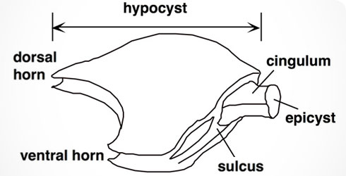
Body plan of Nannoceratopsis.
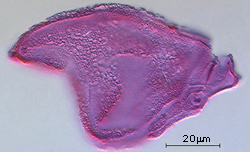
Morphological characters for identifying and classifying fossil dinoflagellate cysts
The most important are:
- the number, shape and arrangement of plates (the tabulation pattern)
- the excystment opening, called the archaeopyle
Right lateral view of Nannoceratopsis gracilis, showing the tabulation details of the sulcus.
Nannoceratopsis tabulation pattern
- The cyst is laterally compressed.
- The 2 large left and right plates build the major part of the cyst and correspond to the hypocyst.
- The epicyst is very small and separated from the hypocyst by the cingulum, or girdle.
- The sulcus is situated at one of the narrow sides and indicates the ventral face. The sulcus is the area where the flagella were inserted in the motile cell.
Most Nannoceratopsis species show 2 antapical horns:
- a ventral horn
- a dorsal horn
The dorsal horn is always longest.
Species are differentiated from each other by:
- their general outline
- the number of horns
N. gracilis has:
- a long dorsal horn
- a reduced ventral horn
N. spiculata is a close relative. It has:
- 2 antapical horns of about equal length
Nannoceratopsis archaeopyle
- Formed by the loss of 1 or more minute plates on the dorsal side of the cingulum.
- This position of the archaeopyle is a unique feature among fossil dinoflagellate cysts.