Hymenolepis microstoma
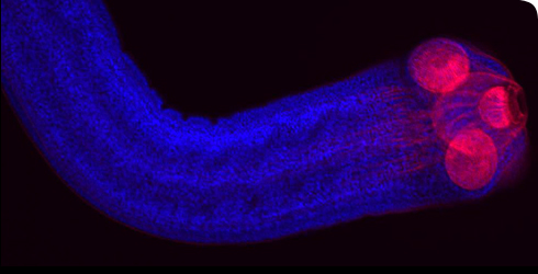
Confocal image of the tapeworm scolex and neck showing the musculature (red) and cell nuclei (blue). © A Gruhl & PD Olson
Hymenolepis microstoma is an enteric (intestinal dwelling) parasite – adult worms live in the bile duct and small intestines of mice, and larvae metamorphose in the haemocoel of beetles.
Species in the genus Hymenolepis (e.g. H. diminuta, H. microstoma, H. nana) have been maintained as laboratory models for studying tapeworm biology since the 1950s.
They are particularly useful for teaching and research purposes as they can be readily maintained in vivo in rodent and beetle hosts. They can also be grown in culture (i.e. in vitro), giving easy manipulation of the life cycle.
Most of our understanding of the basic biology of tapeworms, such as their anatomy, physiology and ultrastructure, stems from work on this genus. Scientists are now characterising the genome and transcriptome of H. microstoma, and thus helping to bring this classical model species into the genomic era.
Species detail
These tapeworms are well known for exhibiting the ‘crowding effect’, which describes the inverse relationship between the number of worms and their size - the larger the infection, the smaller the individual worms, and vice versa.
This means that the overall biomass of the parasite in the host remains constant despite the number of worms present, and may have evolved by preventing undue harm to the host in cases of heavy infection.
We do not know what factors enable the tapeworms to detect and alter their growth in response to their level of ‘crowding’ in the host.
-

Taxonomy
The parasitic tapeworms of mammals, such as H. microstoma, evolved from aquatic tapeworm ancestors that parasitized fish. Discover more about H. microstoma and other flatworm parasites.
-

Distribution and habitat
Hymenolepis microstoma is an obligate parasite that uses mice and beetles in its complex life cycle. It has no mouth or intestine and absorbs nutrients directly from the host’s gut.
-

Biology
Flour beetles are the intermediate host for H. microstoma and become infected when they ingest eggs from infected mouse droppings. Explore the life-cycle of this intestinal parasite and discover how it produces new organs as it grows.
-

Physiology
Find out about the cytology and molecular biology of Hymenolepis microstoma.
-

Lifecycle
Beetles often act as first intermediate hosts for Hymenolepis microstoma, ingesting eggs through mice faeces. Learn more about the lifecycle of this species.
-

Reproduction
Learn how worms reproduce and how their eggs are dispersed.
-

References
Get reference material for Hymenolepis microstoma.
-
Behaviour
Find out about the various hosts of this enteric parasite.
-
Conservation
There are no management practices for this species due to it not affecting Man or domesticated animals. Find out more.
-
Use
Read about the uses for Hymenolepis microstoma and its genus and how these are helping us to understand tapeworm biology.
Images

Light microscopial images of Hymenolepis microstoma showing different regions of the body.
© L Cunningham & PD Olson
Sperm (center) and thin-shelled eggs (left top and bottom) inside a segment of Hymenolepis microstoma.
© L Cunningham & PD Olson
A diagram showing the lifecycle of Hymenolepis microstoma.

Eggs seen inside the segments of Hymenolepis microstoma.
© L Cunningham & PD Olson
Hymenolepis microstoma confocal images (ie. coloured) of the tapeworm scolex and neck showjng the musculature (red or green) and cell nuclei (blue).
© A Gruhl & PD Olson


A word from the author
'The study of Hymenolepis has contributed immensely to our knowledge of tapeworms and the diseases that they cause. By characterizing the genome of this animal, we are helping to bring a ‘classical’ model into the genomic era, and these data are already helping to elucidate the genes that underpin their unique adaptations to parasitism, such as the evolution of a segmented body plan.'
Toolbox
Share this
Glossary
Haemocoel the series of spaces between organs through which haemolymph circulates in arthropods such as beetles.