Taxonomy
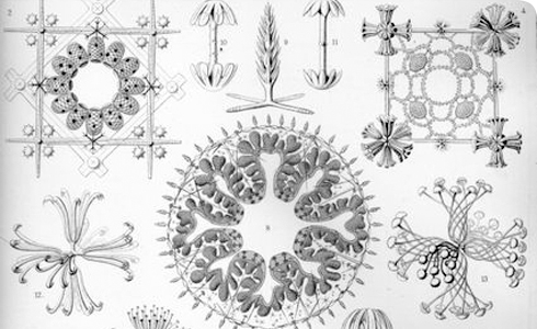
An illustration of Hexactinellae by Haeckel.
Morphology
The body structure of these animals is a thin-walled, cylindrical, vase-shaped tube with a large central atrium. The body is composed entirely of silica in the form of 6-pointed siliceous spicules, which is why they are commonly known as glass sponges. The spicules are composed of 3 perpendicular rays giving them 6 points. Spicules are microscopic, pin-like structures within the sponge’s tissues that provide structural support for the sponge. It is the combination of spicule forms within a sponge’s tissues that helps identify the species.
In the case of glass sponges the spicules 'weave' together to form a very fine mesh which gives the sponge’s body a rigidity not found in other sponge species and allows glass sponges to survive at great depths in the water column. Overlying the spicule framework there is more siliceous tissue called a syncytium which forms very fine fibres which look rather like a cobweb over the framework. The syncytium means that the sponge has a mass of tissue with no defined cell boundaries but lots of nuclei.
The top end of the sponge has a sieve-like disc over the end and the sponge is anchored to the substrate by means of fine, hair-like fibres. These fibres are between 50 and 175mm long. Recent research has proved that these fibres have the same composition as fibre-optical cables like those used in modern telecommunications. They can trap and transmit light. There are several theories about why the sponge does this. One is to attract symbiotic algae or as an attractant for the shrimp which lives within the sponge’s body cavity. Neither of these has been proved and it may just be a coincidental by-product of the spicules’ purity.
While alive, the tissue of these sponges is white to creamy yellow, depending upon how much particulate material they have accumulated in their tissues. The dry 'skeleton' seen in museum collections has often been bleached to create the pure white colour.
New sub species
In 2008, Dr Konstantin Tabachnick, a well regarded researcher on this sponge group, and his co-authors, designated 4 new subspecies of Euplectella aspergillum:
- E. aspergillum regalis (formerly E. regalis Schulze),
- E. aspergillum australicum
- E. aspergillum indonesicum
- E. aspergillum aspergillum.
These were all collected from around the coast of Australia.
Some researchers consider the Hexactinellida sufficiently different from the other sponge groups to require their own Phylum - Sympagla
Etymology
Porifera (Latin): porus = pores; ferre, to bear.
Hexactinellida (Greek): hex = six; aktis, ray.
Syncytium (Greek): syn = with; kytos = cell, hence a multinucleate mass of protoplasm, formed by the merging of cells.
Toolbox
Museum specimen in focus
Registration number: BMNH 1894.8.2.1 Holotype
Glossary
Conchologists is a person who studies the branch of zoology that deals with the study of molluscs, specifically their shells.
Type specimen is a specimen selected to serve as a reference point when a species is first named. As a result, these specimens are extremely important to scientists who are attempting to determine the correct application of a name.
Holotype is the single specimen which is used to define the characteristics for a new species by an author and is therefore designated as the type specimen by the author at the time of publication of that description.