Trichopodiella faurei
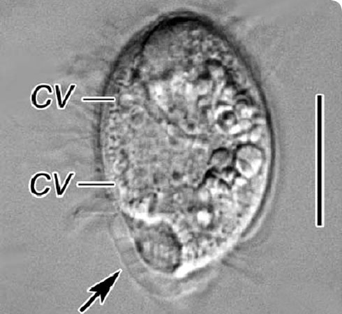
An interference-contrast light microscope image of Trichopodiella faurei.
CV: contractile vacuole.
The arrow points to the cortical alveoli - small sacks in the outer skin.
The line on the right is 0.02mm (for comparison, human hair varies in width between 0.02 and 0.2mm). © J Gong, 2008
Trichopodiella faurei is a marine ciliated protozoan recently described by Museum scientists. It is about 50µm long, equivalent to half the diameter of human head hair.
It has been found free-swimming or attached to solid objects in the Yellow Sea and South China Sea.
The cells are covered with a layer of transparent gel which is foam-like in the posterior region. It also secretes a thread-like (filiform) substance that anchors the cell to a solid base.
This tiny creature was first described in 2008 by Museum researchers.
Species detail
Trichopodiella faurei is a Phyllopharyngean, which means that it has a flattened basket-like structure behind its mouth. (Phyllo - from the Greek for leaf, and pharynx meaning windpipe or throat.)
Living Trichopodiella faurei cells are about 30–60 by 15-30µm. The body is oval, with width slightly greater than the dorsoventral thickness.
-

Taxonomy and distribution
Find out what detailed molecular and microscopic studies of Trichopodiella faurei tell us about protozoa.
-

Evolution and look-alikes
Find out about the evolutionary history of the genus Trichopodiella.
-

References
Get reference material for Trichopodiella faurei.
Images

Trichopodiella faurei from the side, drawn from life.
The body surface is covered with a layer of transparent gel substance which is 1-2µm in thickness, mostly relatively even and smooth but distinctly foam-like in the posterior region of cell, forming cortical alveoli.
The endoplasm is transparent, with numerous 5µm-sized granules and oil droplets. There are usually 2 (seldom 3) contractile vacuoles, each about 5µm in diameter, positioned on the right ventral side.
The arrow indicates small sacks (alveoli) in the outer skin (cortex). The cell nucleus is the large body in the middle of the cell. The mouth is the slit at the top of the drawing supported by stiff rods (trichites). The line on the right is 0.02mm (for comparison, human hair varies in width between 0.02 and 0.2mm.)
The anterior portion is bluntly rounded and slightly wider than the posterior.
© J Gong, 2008
The swimming behaviour of Trichopodiella faurei (top left) and 2 mechanisms of attaching to nearby objects.
© J Gong, 2008
Trichopodiella faurei after staining with silver, which reveals the roots of the cilia - fine hair-like structures with which the cell swims and catches food.
Each dot represents a basal body - a complicated structure that supports and anchors the cilium in the cortex and from which the cilium extends.
CR: caudal ring - a line of cilia around the posterior end of the cell.
Cy: cyrtos - the equivalent of the throat in multicellular animals.
EF: equatorial fragment - a dense collection of basal bodies.
LK and RK: left and right-kineties - a kinety being a row of basal bodies, and left and right are defined as though you were inside the cell.
PK: perioral kinety - the row of cilia immediately to the left of the mouth.
TS: transpodial segment - short kineties that give rise to a tuft of cilia at the posterior end of the cell.
TF: terminal fragment - a dense collection of basal bodies near the front of the cell.
Ma: macronucleus - ciliates have two types of nucleus, a large amorphous body that is active in the cell and a micronucleus that is compact and only active in sexual reproduction.
The line on the left is 0.02mm (for comparison, human hair varies in width between 0.02 and 0.2mm).
© J Gong, 2008
An interference-contrast light microscope image of Trichopodiella faurei.
The body surface is covered with a layer of transparent gel substance which is 1–2µm in thickness, mostly relatively even and smooth but distinctly foam-like in the posterior region of cell, forming cortical alveoli.
The endoplasm is transparent, with numerous 5µm-sized granules and oil droplets. There are usually 2 (seldom 3) contractile vacuoles, each about 5µm in diameter, positioned on the right ventral side.
CV: contractile vacuole - the organelle that can be seen pulsing in life as it expels excess water from the cell.
The arrow points to the cortical alveoli - small sacks in the outer skin.
The line on the right is 0.02mm (for comparison, human hair varies in width between 0.02 and 0.2mm).
The anterior portion is bluntly rounded and slightly wider than the posterior.
© J Gong, 2008
An interference contrast light microscope image of Trichopodiella faurei focussed in the middle of the cell.
The endoplasm is transparent, with numerous 5µm-sized granules and oil droplets. There are usually 2 (seldom 3) contractile vacuoles, each about 5µm in diameter, positioned on the right ventral side.
CV: contractile vacuole - the organelle that can be seen pulsing in life as it expels excess water.
The arrow indicates the posterior depression.
The arrowhead indicates the fairly thin gel-like covering on the front of the cell.
The line on the left is 0.02mm (for comparison, human hair varies in width between 0.02 and 0.2mm).
© J Gong, 2008
An interference contrast light microscope image of the rear portion of Trichopodiella faurei to show the cortical alveoli - small sacks in the outer skin - in the characteristic hexagonal pattern.
The surface of the cortical alveoli exhibits a polygonal pattern.
The line on the right is 0.01mm (for comparison, human hair varies in width between 0.02 and 0.2mm).
© J Gong, 2008Author

$linktitle
A microbiologist specialising in ciliated protozoa. The problems of ciliate nomenclature led to Dave's involvement in the influential Nomencurator project and indirectly to running the EU project Unifying revisionary Taxonomy on the Web.