Taxonomy and distribution
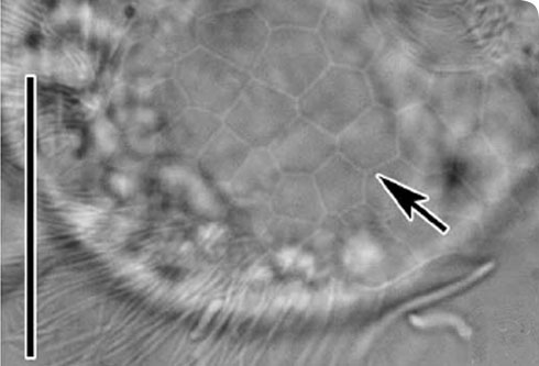
An interference contrast light microscope image of the rear portion of Trichopodiella faurei to show the cortical alveoli - small sacks in the outer skin - in the characteristic hexagonal pattern.
The line on the left is 0.01mm (for comparison, human hair varies in width between 0.02 and 0.2mm). © J Gong, 2008
Morphology
Trichopodiella faurei cells are about 30–60 x 15-30 µm in vivo. The body is oval, with width slightly greater than the dorsoventral thickness.
The anterior portion is bluntly rounded and slightly wider than the posterior.
The body surface is covered with a layer of transparent gel substance which is 1–2µm in thickness, mostly relatively even and smooth but distinctly foam-like in the posterior region of cell, forming cortical alveoli. The surface of the cortical alveoli exhibits a polygonal pattern.
The cytostome is located at about the anterior 1/6 of body length, slit-like, about 10µm long.
The endoplasm is transparent, with numerous 5µm-sized granules and oil droplets. There are usually 2 (seldom 3) contractile vacuoles, each about 5µm in diameter, positioned on the right ventral side.
Trichopodiella faurei produces mucous threads which are used to anchor the cell onto a substrate. This indicates the existence of a glandule, which have been observed in other Trichopodiella species.
The mucous threads emerge ventrally from a conspicuous depression with a collar-shaped border in the posterior portion of cell and are up to 2.5 times body length.
There is no distinct podite. Cilia are about 8–10µm long in vivo.
Diagnostic description
Marine Trichopodiella are oval and approximately 30–60 by 15–30µm in vivo.
They are covered with a layer of transparent gel which is foam-like in the posterior region.
The glandular region is collar-like, conspicuously depressed, and secretes a filiform substance for anchoring the cell to substratum.
Single-rowed perioral kinety are longitudinally oriented, separating left and right somatic kineties.
There are 31–39 somatic kineties, including about 13–19 rows left of the perioral kinety.
There are 2 or 3 contractile vacuoles.
Distribution
Most ciliates are cosmopolitan.
Trichopodiella faurei was first described in 2008, and so far two populations of have been found in:
- coastal waters of the Yellow Sea, Qingdao (36° 08' N; 120° 43' E), northern China
- Daya Bay (22°42'N 114°31'E), South China Sea, southern China